Veterinary Radiology Interpretation Services for Veterinary Practices
Diagnostic imaging can provide valuable information about a patient, but understanding what those images show requires experience and clinical context. AxisVet provides veterinary radiology interpretation services that give veterinary practices access to board-certified radiologists for expert review of diagnostic imaging studies.
Our radiologists evaluate imaging findings in the context of the clinical information provided by your team, helping you better understand abnormalities and their potential significance. With specialist interpretation and collaborative support, AxisVet helps veterinarians incorporate imaging findings into the broader diagnostic process and make informed decisions for their patients.
Why Does Veterinary Radiology Interpretation Matter
Veterinary radiology requires a detailed understanding of anatomy, disease patterns, imaging techniques, and the differences that can occur across species, breeds, and stages of life. Identifying an abnormality is only part of the process. Radiologists also consider its location, appearance, distribution, and relationships to other findings within the study and patient history.
Board-certified veterinary radiologists bring specialized training and extensive experience in diagnostic imaging to this process. Their expertise can be particularly valuable when findings are subtle, unexpected, complex, or difficult to interpret.
Specialist interpretation also provides another clinical perspective for your team. The radiologist evaluates the imaging study and communicates relevant findings, while the veterinarian managing the patient considers that information alongside the physical examination, medical history, laboratory results, and other diagnostics.
In this way, AxisVet’s radiologists serve as an extension of your clinical team, providing imaging expertise that supports rather than replaces your veterinary judgment.
What Veterinary Radiologists Evaluate
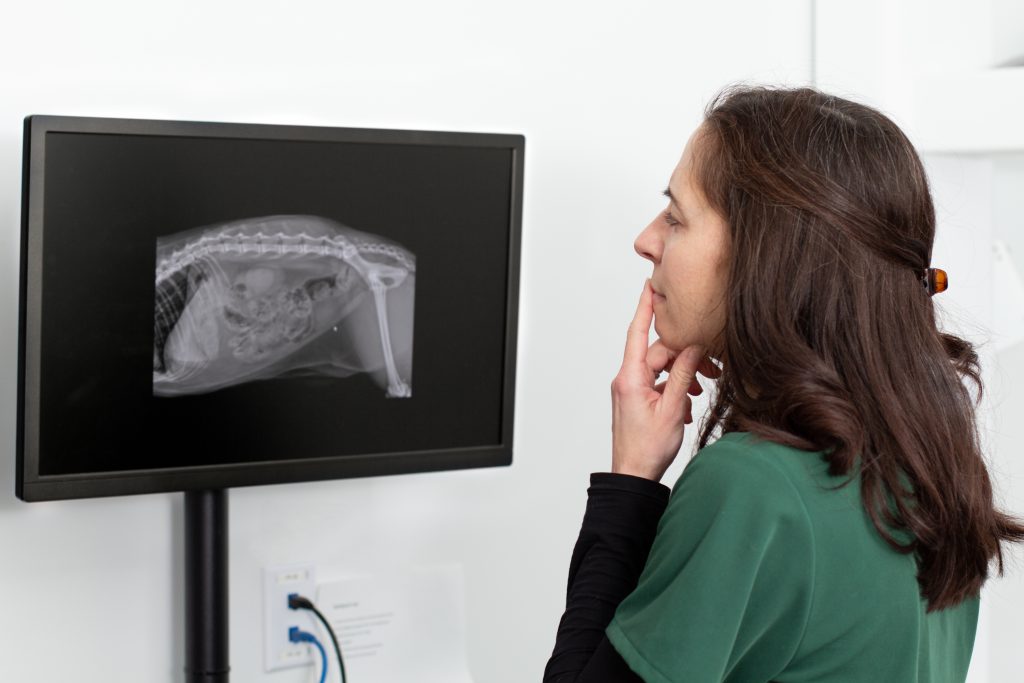
Veterinary radiology interpretation follows a systematic approach to evaluating an imaging study. Rather than focusing only on the suspected problem, a radiologist reviews the available images for relevant abnormalities and considers how individual findings may relate to one another.
Depending on the study and area being evaluated, a veterinary radiologist may assess:
- Organ size, shape, position, and appearance
- Skeletal structures and joints
- Thoracic and pulmonary patterns
- Abdominal structures and soft tissues
- The location and distribution of identified abnormalities
Radiologists must also distinguish meaningful abnormalities from normal variations. Patient factors such as species and age can influence how structures appear, while image positioning and quality may affect what can be evaluated.
The radiologist then considers these visual findings alongside the clinical history and reason for the study. This combination helps place abnormalities in context and allows the report to provide useful information to the veterinarian managing the case.
Diagnostic Imaging Modalities Interpreted by AxisVet
Different diagnostic imaging modalities provide different types of information to veterinary professionals. AxisVet’s board-certified veterinary radiologists interpret radiographs, ultrasound, fluoroscopy, CT, and MRI studies, providing specialist expertise across routine and advanced imaging cases.
Radiograph Interpretation
Radiographs are commonly used to evaluate the thorax, abdomen, bones, joints, and other anatomical structures. During radiographic interpretation, a veterinary radiologist may assess changes in bone and joint structures, organ size or position, pulmonary patterns, and other findings relevant to the area being examined.
The radiologist also considers image quality and positioning, which can affect what is visible on a radiographic examination. These findings are then considered alongside the clinical information provided by the submitting veterinarian.
Ultrasound and Fluoroscopy Interpretation
Ultrasound provides detailed information about soft tissues and internal organs. A veterinary radiologist evaluates the appearance and architecture of these structures and identifies abnormalities that may be relevant to the patient’s clinical signs.
Fluoroscopy provides dynamic imaging that allows certain structures and processes to be observed in motion, helping the radiologist evaluate functional findings and their clinical significance.
CT and MRI Interpretation
CT and MRI provide detailed cross-sectional images that can be valuable when evaluating complex anatomical areas or conditions that require advanced diagnostic imaging.
These studies can generate more enhanced imaging capabilities. A veterinary radiologist evaluates the location, extent, and characteristics of relevant findings to provide a focused interpretation of the study.
How a Detailed Clinical History Supports Radiology Interpretation
Images tell an important part of the story, but relevant clinical information helps the radiologist understand the questions your veterinary team is trying to answer.
Relevant clinical information may include:
- The reason for the imaging study
- The presenting complaint and relevant patient history
- Examination findings
- Relevant diagnostic results
- Specific clinical questions your team wants the radiologist to address
For example, knowing whether an abnormality is an unexpected finding or relates to an established clinical concern can influence how the radiologist approaches the study and communicates its significance.
Focus on information that is relevant to the current imaging study. This context can help the radiologist provide an interpretation that is useful to the veterinarian managing the case.
What to Expect from a Veterinary Radiology Report
A teleradiology report outlines the specialist’s interpretation of the submitted study. It should give the veterinarian a clear understanding of the relevant findings and their potential significance within the limits of the available images and clinical information.
The report may also provide an overall impression of the relevant findings. When appropriate, the radiologist may also recommend additional imaging, diagnostics, monitoring, or specialist evaluation that could provide further information.
These recommendations are intended to support the next stage of the diagnostic process rather than dictate patient management. The submitting veterinarian can consider the interpretation alongside the patient’s history, examination findings, diagnostic results, and response to treatment when determining the appropriate next steps.
AxisVet also understands that timely information matters when veterinarians are making clinical decisions. Veterinary practices can access standard reports within hours, with STAT reporting available in under an hour when urgent interpretation is needed.
Collaboration with Board-Certified Veterinary Radiologists
AxisVet’s veterinary radiology interpretation services are backed by board-certified veterinary radiologists with the expertise to evaluate both routine and complex diagnostic imaging studies.
When a report raises questions or a finding requires clarification, AxisVet gives veterinary teams direct access to radiologists for further discussion. This allows veterinarians to better understand imaging findings, recommendations, or other aspects of the interpretation that may affect the next stage of a patient’s workup.
This collaborative support is central to AxisVet’s interactive approach. Technology makes remote image interpretation efficient and accessible, while efficient access to our radiologists remains the center of our customer experience.
Understanding the Scope and Limitations of Imaging Interpretation
Diagnostic imaging is an important part of veterinary medicine, but an imaging study represents one component of a patient’s overall clinical evaluation.
Some abnormalities have characteristic imaging appearances, while other findings may be less specific or overlap with multiple potential conditions. Technical factors, patient positioning, the type of imaging performed, and the structures included in the study can also influence what the radiologist is able to evaluate.
When imaging findings alone cannot answer the clinical question, the radiologist may recommend considering other clinical information, additional diagnostics, or additional specialist support such as a veterinary internal medicine consultation when appropriate.
The veterinarian managing the patient remains responsible for bringing these pieces together. Radiology interpretation provides specialist insight into the imaging study, while the patient’s history, physical examination, laboratory findings, other diagnostics, and clinical response provide additional information needed to understand the complete case.
Frequently Asked Questions About Veterinary Radiology Interpretation
What clinical information should I provide for a radiology interpretation?
Provide information that is relevant to the imaging study, including the reason for the examination, presenting complaint, relevant patient history, examination findings, and diagnostic results. Including specific clinical questions can also help the radiologist understand what your team is evaluating.
What types of veterinary imaging can AxisVet radiologists interpret?
AxisVet provides specialist interpretation of radiographs, ultrasound, fluoroscopy, CT, and MRI studies. These modalities can provide different types of diagnostic information depending on the patient and clinical concern.
Can I discuss imaging findings with the reporting radiologist?
Yes. Direct access to specialists is an important part of AxisVet’s approach. Veterinary teams can communicate with radiologists when additional discussion or clarification about imaging findings or recommendations is needed.
What is included in a veterinary radiology interpretation?
A radiology interpretation communicates relevant findings from the submitted study and an overall assessment of their significance. Depending on the case, the radiologist may also provide recommendations for additional imaging, diagnostics, monitoring, or specialist input when appropriate.
When might additional diagnostic imaging be recommended?
Additional imaging may be recommended when the initial study does not fully answer the clinical question or when a finding would benefit from further evaluation. The appropriate next step depends on the imaging findings, available clinical information, and needs of the individual patient.
Contact AxisVet for Veterinary Radiology Interpretation
AxisVet gives veterinary practices access to board-certified radiologists who provide clinically relevant imaging interpretation and collaborative specialist support. Whether your team needs interpretation of routine radiographs or advanced diagnostic imaging, our radiologists provide the expertise and context needed to help you evaluate your cases.