Why More Veterinarians Are Turning to Telemedicine
Telemedicine adoption is rising because today’s challenges require more than in-person staffing can supply. Many practices deal with limited access to specialists, lengthy turnaround times from national providers, and day-to-day workflow bottlenecks that slow clinical decisions. Modern veterinary telemedicine addresses these issues by giving practices access to specialists who deliver timely reads and structured interpretations that fit directly into the team’s workflow.
Independent practices gain access to radiologists, cardiologists, and internal medicine support without adding new employees. Larger groups benefit from predictable remote diagnostics for vets across all locations. The result is higher throughput, fewer backlogs, and a more organized workday.
At AxisVet, our goal is to help clinics experience these operational gains by removing the uncertainty that often surrounds remote diagnostics.
What Veterinary Telemedicine Includes Today
Telemedicine covers a wide spectrum of diagnostic and clinical support services. Each one plays a distinct role in helping teams work more efficiently, giving clinicians actionable reports and guidance that move cases forward.
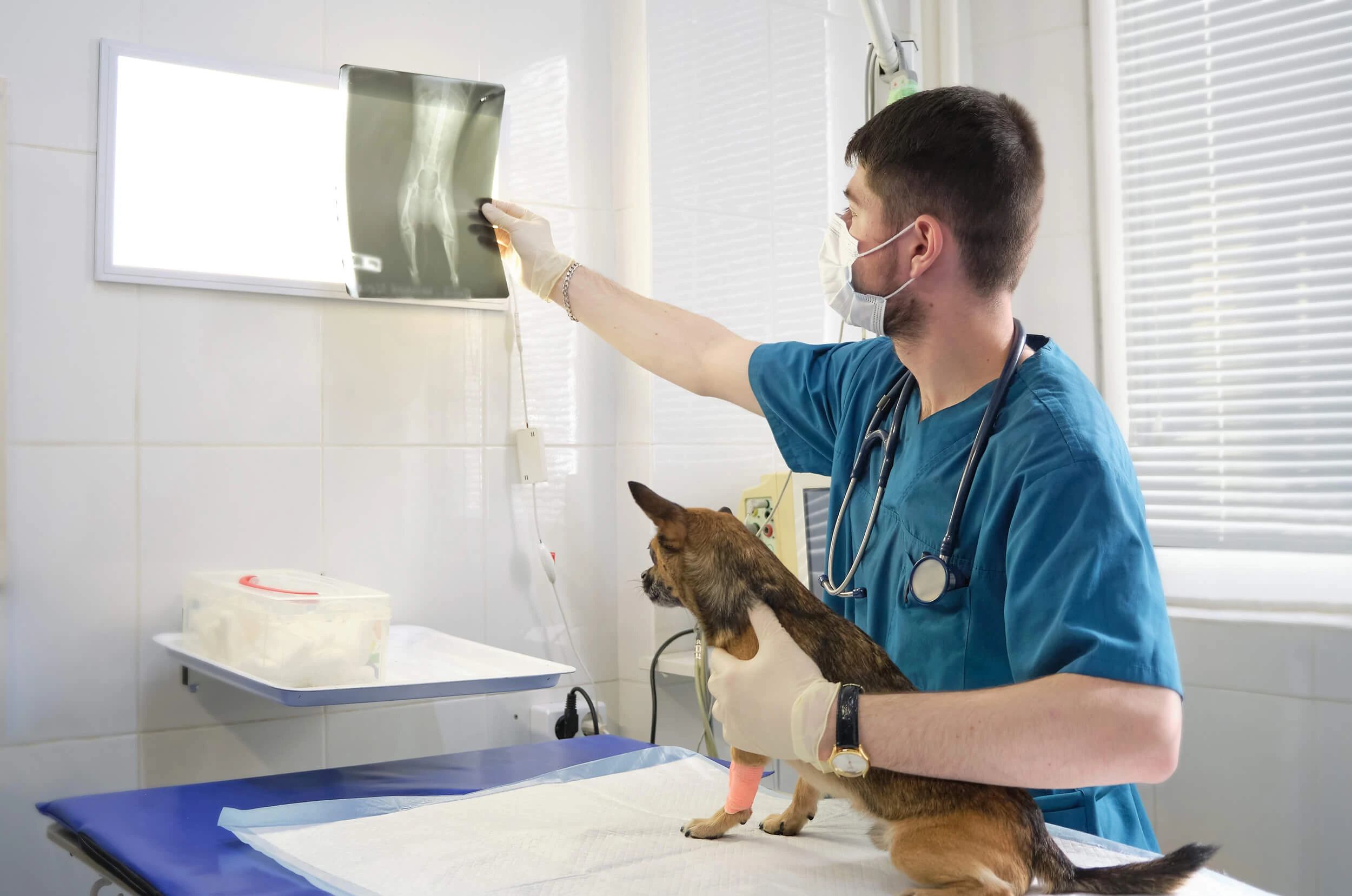
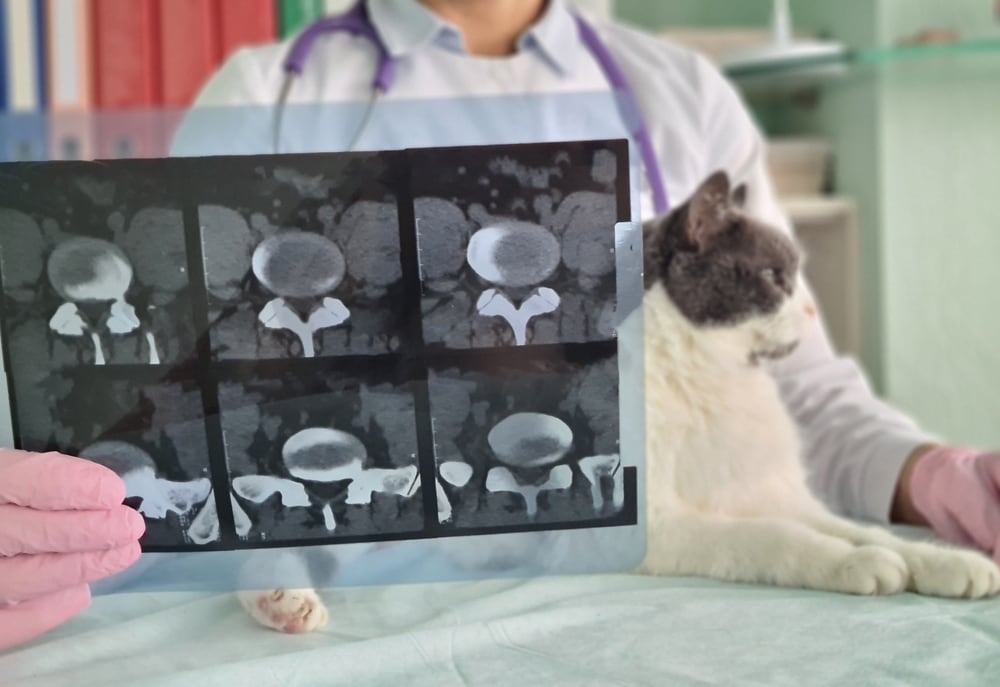
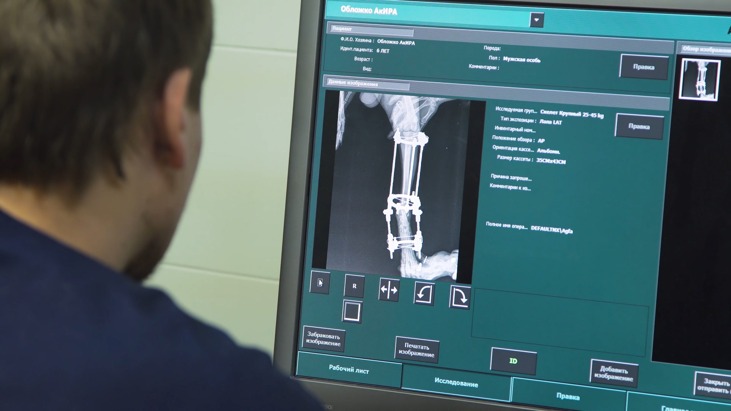
- Teleradiology services: High-volume interpretation of imaging studies, handled by board-certified radiologists. Clinics can submit cases quickly and receive structured reports that support timely decisions.
- Telecardiology for veterinarians: Interpretation of cardiac studies, rhythm evaluations, congenital concerns, and chronic disease cases handled by trained cardiology specialists.
- ECG and arrhythmia evaluations: Analysis of rhythm abnormalities to help clinicians identify the cause of syncope, collapse, murmurs, or exercise intolerance.
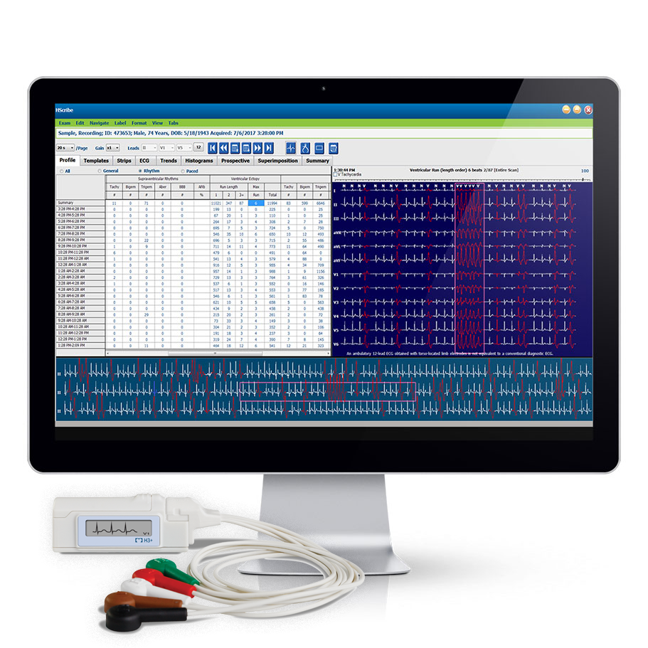
- Holter monitoring services: Continuous cardiac monitoring used for arrhythmia detection and long-term case management.
- Internal medicine consultations: Case support for complex medical conditions, chronic disease, and unusual presentations that require a specialist’s perspective.
How Telemedicine Improves Veterinary Workflow Efficiency
Many daily bottlenecks in veterinary practice trace back to slow diagnostics. When read times lag, the entire team waits. Patients remain in limbo, follow-up calls pile up, and staff lose time troubleshooting incomplete studies or resubmitting images. Telemedicine smooths this process by connecting clinics with specialists who deliver prompt, structured reports.
Fast read times support same-day decisions, which reduces patient wait periods. Clear interpretations reduce repeated questions and eliminate unnecessary back-and-forth communication. Technicians also gain time because they spend less of the day chasing image resubmissions or confirming uploads.
Below is a quick look at a traditional workflow vs. telemedicine-enhanced workflow.
Traditional Workflow
- Veterinary clinic sends images to a national service
- Delayed turnaround stalls treatment
- Team revisits incomplete details
- Client communication becomes fragmented
- Procedures shift to another day due to slow reads
Telemedicine-Enhanced Workflow
- Prompt submission to AxisVet specialists
- High-quality reports support same-day planning
- Fewer reworks or clarifications
- Clients receive faster updates
- Teams schedule procedures with more consistency
Teleradiology’s Role in Better Case Management
Radiology drives a large portion of diagnostic volume in general practice, which means delays in imaging interpretation can disrupt an entire day of appointments. Reliable read times help clinics plan procedures, schedule callbacks, and communicate with clients more effectively. High-quality reports also shape surgical decisions, rule-outs, and referral planning.
At AxisVet, our teleradiology services team focuses on clarity and consistency. Reports follow a structured format that reduces miscommunication among veterinarians and technicians. Radiologists remain accessible for follow-up questions so teams can resolve uncertainties quickly. These steps support more efficient case management and a better diagnostic experience for customers who expect timely information and smoother service.
What Makes Teleradiology Effective
- Reliable turnaround time that aligns with clinic workflow
- Clear formatting to reduce confusion
- Access to specialists for case discussion
- Reports that support patient-side decisions on the same day
These elements create more predictable operations and a more organized clinical environment.
Telecardiology and Holter Services for Busy Clinics
Cardiology remains one of the most difficult specialties for clinics to access. Referral wait times are often long, and many regions lack accessible cardiologists. Telehealth service solves this gap by connecting veterinarians with specialists who support rhythm evaluations, murmur workups, and long-term disease management.
Our telecardiology team helps clinics triage and manage cardiac concerns without delay. Clear, actionable reports guide medication adjustments and follow-up planning. Holter monitoring gives veterinarians a practical way to assess intermittent arrhythmias and long-term rhythm patterns without sending patients away from the practice.
Key benefits of telecardiology support include:
- Clear guidance for acute and chronic cardiac cases
- Arrhythmia detection through Holter and ECG interpretation
- Structured reporting that supports next-step decisions
- Fewer unnecessary referrals, leading to higher client satisfaction
These services provide faster image interpretation and more organized case management, helping teams keep control of their day.
Why Telemedicine Works for Independent and Multi-Location Clinics
Independent clinics face staffing limitations that make daily operations challenging. Hiring on-site radiologists or cardiologists is rarely practical. Telemedicine helps these practices match the capabilities of larger hospital groups without expanding headcount. Reliable turnaround times also reduce operational strain created by slow national diagnostic queues.
Multi-location groups benefit in other ways. Clinical quality becomes consistent across all sites, and managers gain predictable diagnostic timelines that support scheduling. Fewer delays mean more complete cases and a smoother experience for both staff and clients. At AxisVet, we focus on communication and reliability so clinics can plan their workflow around steady diagnostic support instead of unpredictable delays.
Choosing the Right Telemedicine Partner
Selecting a telemedicine service partner is a strategic decision for any clinic. Report clarity, access to specialists, turnaround times, pricing structure, and communication style all influence how smoothly the partnership functions. Clinics should also consider how well a provider supports their preferred workflow and how responsive the team is when questions arise.
AxisVet compares favorably to national providers whose delays often interrupt case progression. We focus on providing consistent timelines, structured reports, and direct communication with specialists. This approach supports better outcomes for both patients and staff.
What to Look For in a Telemedicine Partner
- Clear report style that fits your clinical approach
- Transparent pricing with no surprise fees
- Predictable turnaround
- Accessible specialists for case discussion
Teams that evaluate these criteria usually find a partner who supports their long-term growth and workflow goals.
Telemedicine That Helps Your Veterinary Team Do More Each Day
Modern telemedicine for veterinarians plays a central role in how clinics organize their day. It contributes to faster decisions, improved diagnostic accuracy, and a more controlled workload for technicians and DVMs. Those advantages help clinics deliver better veterinary care and a more consistent experience for clients.
AxisVet provides veterinary telehealth consultations designed to support daily operations and reduce the strain caused by limited specialist access. To learn more about how our services can support your workflow, contact AxisVet today.


