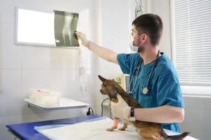
Interpreting heart health in cats can be complex, particularly when signs are subtle or patients are struggling to breathe. With thoughtful technique and the right tools, radiographs remain an essential part of cardiac evaluation. Below, we share five practical tips to help you use X-rays more effectively in feline cardiac cases—especially when time and patient tolerance are limited. These insights can support earlier diagnosis, guide next steps, and improve outcomes for even your most delicate patients.
1. How Do You Balance Safety and Image Quality During X-Rays?
Cats with heart conditions often present late in the disease process, with respiratory distress or vague clinical signs. In these situations, capturing the ideal X-ray views may not be safe. A single lateral view can often provide valuable information without overhandling the cat. Use clinical judgment to determine when additional images are appropriate. If the cat stabilizes, consider light sedation to safely acquire orthogonal views and improve positioning. Patient comfort should always guide the process—after all, the best images are ones you can obtain without causing harm.
2. What Should You Look for When Evaluating the Heart Shape?
In a healthy cat, the cardiac silhouette typically resembles an elongated football on lateral radiographs. Subtle changes in this silhouette can be early indicators of structural heart disease. For example, widening of the cranial aspect of the heart may suggest left atrial enlargement—a common finding in conditions like hypertrophic cardiomyopathy. In some cases, the heart may even take on a Valentine-shaped appearance. Being familiar with these changes helps you recognize abnormal patterns quickly and improves the likelihood of an accurate diagnosis.
3. When and Why Should You Add an Echocardiogram?
While X-rays provide valuable information about cardiac size and pulmonary structures, some diseases can go undetected without further diagnostic imaging. An echocardiogram allows for real-time assessment of myocardial thickness, chamber function, and the presence of thrombi—often before radiographic changes appear. This makes it a critical tool when ruling in or out primary cardiac disease. If you suspect heart disease but aren’t seeing definitive signs on radiographs, ultrasound can clarify the picture and help you intervene earlier.
4. How Do Age-Related Changes Affect Heart X-Rays?
Geriatric cats often present with incidental radiographic findings that can mimic pathology. Age-related changes like vertebral deviation, mild tracheal elevation, or apparent cardiac rounding can resemble disease. Knowing what’s expected in older patients helps prevent overinterpreting benign features as pathology. It’s also important to consider concurrent conditions like chronic kidney disease or hyperthyroidism, which can influence both the heart’s appearance and function. Context matters—and comparing current films to past studies is often helpful.
5. When Should You Seek a Second Opinion?
Interpreting feline thoracic radiographs—especially in complex cardiac cases—can be challenging. If you’re unsure if changes are pathologic or age-related, or if cat radiographs are inconclusive, it’s always worth reaching out. At AxisVet, we offer teleconsultation services that connect you with experienced specialists who can provide fast, trusted feedback. Our services help clarify difficult cases, guide further testing, and support confident clinical decisions—so your patients get the care they need, when they need it.
Partner With AxisVet for Expert Support
Radiographs are a powerful diagnostic tool for assessing feline heart health—but they’re even more effective when paired with collaborative care. At AxisVet, our veterinary diagnostic imaging services are designed to support you at every step, from case triage to expert image review. Whether you’re managing a stable chronic condition or triaging an acute respiratory crisis, our team is here to help.
Contact us to learn more about what we have to offer, including veterinary radiology reviews, echocardiogram interpretation, and second opinions tailored to your veterinary medicine practice’s needs.